Machine-Learning-Assisted Microscopy : From Smart Scanning Approaches to the Generation of Synthetic Super-Resolution Images
Super-resolution microscopy (or optical nanoscopy) techniques allow the characterization of molecular interactions inside living cells with unprecedented spatiotemporal resolution. These techniques come with several layers of complexity in their implementation. My research team focuses on transdisciplinary approaches at the interface of molecular neurosciences, multimodal optical nanoscopy, and machine learning to study structure/function relationship of synapses in the brain.
Date
- 10 février 2020
Heure
13h30 à 14h30
Localisation
Université Laval
Pavillon Adrien-Pouliot
Local 3370
Coûts
Gratuit
Super-resolution microscopy (or optical nanoscopy) techniques allow the characterization of molecular interactions inside living cells with unprecedented spatiotemporal resolution. These techniques come with several layers of complexity in their implementation. My research team focuses on transdisciplinary approaches at the interface of molecular neurosciences, multimodal optical nanoscopy, and machine learning to study structure/function relationship of synapses in the brain.
We develop machine learning and deep learning tools to increase the adaptability and accessibility of high-end imaging methods (e.g. optical nanoscopy) to complex experimental paradigms. Recently, we implemented a machine learning assisted optimization framework for optical nanoscopy allowing real-time optimization of multi-modal live-cell imaging of synaptic activity and structure proteins. We also implemented diverse deep learning approaches for high throughput microscopy image analysis, allowing us to characterize activity-dependent remodelling of neuronal proteins. We develop weakly supervised deep learning strategies to reduce the burden of extensive labeling of complex images and evaluate how they can be applied to real-time microscopy image analysis.
We aim at developing new AI-assisted microscopy techniques that will adapt in real-time to the sample, predict changes in the structures and modify the experimental protocol depending on the measured response to a stimuli.
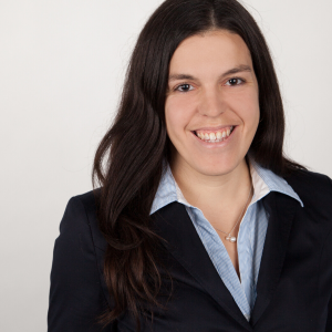
Conférencière
Professeure adjointe, Faculté de médecine, Université Laval
Titulaire, Chaire de recherche du Canada en nanoscopie intelligente de la plasticité cellulaire
Responsable de l’axe Santé et étude du vivant, IID
Flavie Lavoie-Cardinal a obtenu son baccalauréat, sa maitrise et son doctorat en chimie de l’Université de Siegen en Allemagne. Elle a ensuite été stagiaire postdoctoral (entre 2011 et 2014) en Biophysique dans le laboratoire du Prof. Stefan W. Hell (prix Nobel de Chimie en 2014) sur le développement de la microscopie de super-résolution. Elle a effectué un deuxième stage postdoctoral dans le laboratoire du Prof. Paul De Koninck en Biophotonique entre 2014 et 2017 sur l’application de la super-résolution aux neurosciences cellulaires et moléculaires. Elle a ensuite été chercheure indépendante au centre CERVO et Professeure associée au département de Physique, Génique Physique et Optique de l’Université Laval.
Mme Cardinal a mis sur pied un programme de recherche qui utilise et développe l’apprentissage automatique et l’apprentissage profond pour la microscopie. Ses projets portent d’une part sur l’amélioration des performances des techniques de microscopie et d’autre part sur le développement de techniques d’analyse quantitatives des images obtenues.
Restons en contact!
Vous souhaitez être informé des nouvelles et activités de l'IID? Abonnez-vous dès maintenant à notre infolettre mensuelle.
